
Atypical Lesions
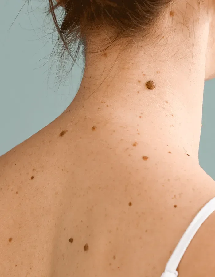
The term atypical lesion (also called an atypical naevus, dysplastic naevus, or atypical mole) refers to a pigmented skin spot or mole that displays certain irregular features, either in its size, shape, colour, or border, that distinguish it from an ordinary benign mole. These features may be detected clinically by the naked eye, with a dermatoscope, or on histopathological examination of a tissue sample.
Most atypical lesions are benign. However, they require careful assessment by a qualified clinician because some features associated with atypicality can also be seen in early melanoma, and individuals with multiple atypical naevi have a higher lifetime risk of developing melanoma than the general population.
This material is intended for general educational purposes and is produced in accordance with the Therapeutic Goods Advertising Code 2021 (Cth). No specific therapeutic goods (including prescription medicines or medical devices) are named or promoted in this document. Treatment suitability is determined only after individual clinical assessment. YourSkin Clinic practitioners are registered with the Australian Health Practitioner Regulation Agency (AHPRA).
Treatments involving prescription medicines may only be recommended and supplied following a valid prescription issued by an authorised prescriber. Not all treatments described will be suitable for all individuals. Results are not guaranteed and vary between patients.
Three major types of Skin Cancer
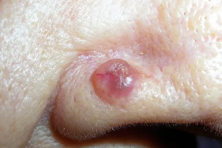
Basal Cell Carcinoma
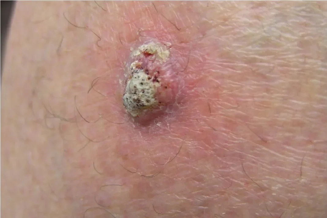
Squamous Cell Carcinoma
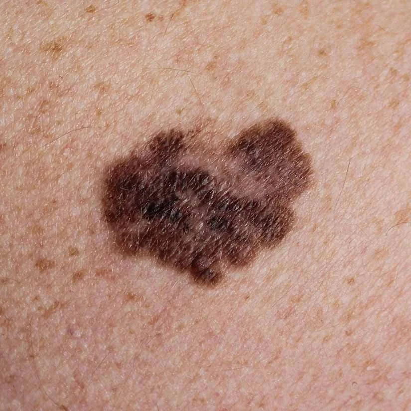
Melanoma
-
The ABCDE criteria are a widely used clinical tool to help identify features of a lesion that warrant further assessment. They are a useful guide for both clinicians and patients, but are not a substitute for professional evaluation. A lesion displaying one or more ABCDE features should be assessed by a qualified clinician promptly.
Asymmetry
One half does not mirror the other
Border
Irregular, ragged, or poorly defined edges
Colour
Multiple shades or uneven pigmentation
Diameter
Greater than 6mm, though smaller lesions also warrant review
Evolution
Any change in size, shape, colour, or new symptoms
-
The development of atypical naevi is influenced by a combination of genetic and environmental factors:
UV exposure: Cumulative sun damage, particularly during childhood and adolescence, and episodes of blistering sunburn, is strongly associated with the development of naevi, including atypical ones
Genetics: A tendency to develop multiple atypical naevi (dysplastic naevus syndrome) can run in families. Certain gene variants, including mutations in CDKN2A and CDK4, significantly increase both naevus burden and melanoma risk
Skin type: Individuals with fair skin, light eyes, and red or blonde hair have less melanin protection and are at greater risk
Immune status: Immunosuppression (from medication or illness) is associated with an increased number and atypicality of skin lesions
High naevus count: Having a large number of ordinary moles (50 or more) is itself a risk factor for developing atypical naevi and melanoma
-
Dysplastic naevus syndrome (also called familial atypical multiple mole melanoma syndrome, or FAMMM) is a condition characterised by:
A large number of naevi (often 50 or more, sometimes hundreds)
At least some of which display atypical features
A personal or family history of melanoma in some definitions
Individuals with this syndrome have a significantly elevated lifetime risk of developing melanoma compared with the general population. This risk is estimated to be 10 times or more higher in some studies. These patients require regular, systematic skin surveillance by an experienced clinician, and may benefit from referral to a dermatologist and genetic counselling if there is a strong family history of melanoma.
-
A thorough skin lesion consultation involves several components:
Medical and skin history: Including personal and family history of skin cancer, previous biopsies or excisions, sun exposure history, medications, and immune status
Naked-eye examination: Visual inspection of the lesion or lesions of concern in good lighting
Dermoscopic examination: Detailed assessment of each lesion using a dermoscope
Total body skin examination (if requested or recommended): A systematic examination of the entire skin surface to identify any additional lesions of concern
Documentation: Clinical photographs of lesions of concern are taken and retained in your medical record for comparison at future visits
Clinical decision: Based on the assessment, the clinician will advise whether the lesion can be monitored, warrants biopsy, or requires excision
You are encouraged to point out any lesion that concerns you, however minor it may seem. Never hesitate to ask your clinician to look at a specific spot. Skin checks are bulk-billed for all pension/DVA card holders.
-
A biopsy is a minor surgical procedure in which a sample of skin tissue (or the entire lesion) is removed and sent to a pathology laboratory for examination under a microscope by a dermatopathologist. Biopsy is the definitive method of diagnosing a skin lesion: clinical and dermoscopic assessment can inform the level of suspicion and guide decision-making, but histopathological analysis provides a tissue-level diagnosis.
A biopsy may be recommended when:
A lesion displays dermoscopic features of concern that cannot be definitively classified as benign
A lesion is changing or has new symptoms (bleeding, crusting, itch)
Clinical assessment cannot exclude malignancy with sufficient confidence
The clinician wishes to confirm that a lesion is benign before committing to monitoring
Being recommended a biopsy does not necessarily mean your doctor suspects cancer. It means they cannot definitively exclude it without tissue analysis, and appropriate management requires certainty.
-
The biopsy technique is selected based on the size, location, and clinical features of the lesion:
Punch biopsy: A circular cutting tool removes a cylindrical core of tissue. Useful for larger lesions where complete excision is not practical or for non-pigmented lesions. However, punch biopsy of a pigmented lesion risks sampling error (missing the most abnormal area), so is generally not the first-line approach for suspected melanoma.
Shave biopsy: The surface of a lesion is shaved off with a blade. Appropriate for some raised lesions; not suitable for suspected melanoma as it does not allow accurate measurement of lesion depth (Breslow thickness), which is essential for staging and management planning.
Incisional biopsy: A portion of a large lesion is removed. Used in specific circumstances where complete excision is not feasible pending a diagnosis.
-
A skin biopsy at YourSkin Clinic is a minor procedure performed under local anaesthetic. The process is as follows:
Local anaesthesia: A small injection of local anaesthetic is administered around the lesion. This causes a brief stinging sensation; the area will then be numb within a minute or two.
Excision: The lesion is removed using a scalpel or punch tool. You will feel pressure but should not feel pain. Let your clinician know immediately if you experience any discomfort.
Closure: Depending on the size and site, the wound may be closed with sutures, adhesive strips, or allowed to heal by secondary intention.
Wound care instructions: Full written aftercare instructions are provided. Most wounds heal within 2 to 3 weeks; sutures are typically removed at 7 to 14 days depending on the site.
Specimen dispatch: The tissue is sent to a certified pathology laboratory. YourSkin Clinic uses Douglass Hanly Moir located in Edgecliff Centre. Results typically take 5 to 10 business days.
-
Pathology results should always be interpreted in the context of your individual clinical presentation by the doctor who assessed you. Do not rely solely on written information to understand your results. A follow-up appointment to discuss findings is essential. Once your results have been received, we will contact you to book an appointment, this can be face-to-face or via telehealth.
-
A biopsy typically involves only minor discomfort because local anesthesia is used to numb the area. While you may feel a sting from the anesthetic injection and pressure during the sample collection, the procedure itself is designed to be manageable, with mild soreness common afterward.
-
Yes, our specialist general practitioner has many years of experience in surgically removing SCC and BCC. The procedure is generally performed in clinic using local anesthetic.