Skin Checks
A skin check is a thorough head-to-toe examination of your skin performed by a doctor. The purpose is to identify any spots, moles, or lesions that may be cancerous, pre-cancerous, or otherwise warrant monitoring or treatment.
Australia has one of the highest rates of skin cancer in the world. Two in three Australians will be diagnosed with skin cancer by the age of 70. The good news is that skin cancers detected early are highly treatable, which is why regular skin checks are one of the most important things you can do for your health.
Who should get a skin check?
Everyone is encouraged to have regular skin checks, but you are at higher risk and should be particularly vigilant if you:
Have fair skin, light eyes, or red or fair hair
Burn easily or have a history of sunburn, particularly in childhood or adolescence
Have a large number of moles (more than 50)
Have a personal or family history of melanoma or other skin cancers
Have had previous skin cancers or pre-cancerous lesions removed
Work or spend significant time outdoors
Have used solarium or sunbed equipment
Are immunosuppressed: for example, following an organ transplant or due to certain medications or medical conditions
Are over 50 years of age, though skin cancer can occur at any age
If you are unsure whether you are due for a check, it is always better to book one and be reassured than to wait.
Before your appointment
Remove nail polish from fingers and toes if possible as melanoma can develop under nails
Tie back or remove hair clips so the scalp can be examined
Avoid applying fake tan as it can obscure lesions
Be prepared to undress to your underwear; a full skin check includes your back, scalp, between the toes, and other areas not routinely visible
What to expect
During the appointment:
Your doctor will systematically examine your entire skin surface, including:
Face, ears, lips, and scalp
Neck, chest, and trunk - both front and back
Arms, hands, fingers, and under the nails
Legs, feet, toes, and soles
A dermatoscope is typically used; this is a handheld magnifying device with a built-in light source that allows the doctor to examine the structure of a lesion beneath the skin surface in detail. This device significantly improves the accuracy of diagnosis compared to the naked eye alone.
After the examination:
Our doctor will discuss their findings, explain any lesions of concern, and recommend a management plan. This may include:
Reassurance and routine monitoring
A short-interval review of specific lesions
A biopsy to confirm or exclude a diagnosis
Referral for surgical removal of a suspicious lesion
Frequently asked questions
-
Regular self-examination between appointments is important. Get to know your skin so you notice what is new or changing. Use the ABCDE guide when assessing moles and spots:
A: Asymmetry: One half of the lesion does not match the other
B: Border: Edges are irregular, ragged, notched, or blurred
C: Colour: Uneven colour, or multiple shades of brown, black, red, white, or blue within the same lesion
D: Diameter: Larger than 6mm (roughly the size of a pencil eraser). Although melanomas can be smaller
E: Evolution: Any change in size, shape, colour, or any new symptom such as bleeding, itching, or crusting
The most important of these is Evolution, any spot that is changing should be assessed promptly, regardless of whether it meets the other criteria.
Also seek review for:
A sore or ulcer that does not heal within 4-6 weeks
A new lesion that is growing quickly
Any spot that bleeds spontaneously or with minimal trauma
Anything that simply looks or feels different from your other spots. Clinicians sometimes call this the "ugly duckling" sign
Perform a self-check monthly in good lighting, using a full-length mirror and a hand mirror for hard-to-see areas. Ask a partner or family member to help examine your scalp and back. Work systematically from head to toe so no area is missed.
Keep a simple record or photograph concerning spots on your phone so you can track whether they change between appointments.
-
Skin cancer is largely preventable. UV radiation is present year-round in Australia. And not just in summer and not only on sunny days. Follow the Cancer Council's Slip, Slop, Slap, Seek, Slide guidelines:
Slip on sun-protective clothing
Slop on SPF 50+ broad-spectrum, water-resistant sunscreen: reapply every 2 hours when outdoors
Slap on a broad-brimmed hat
Seek shade, particularly between 10am and 3pm when UV is strongest
Slide on wrap-around sunglasses
Sunscreen should be worn daily on exposed areas year-round (not just at the beach). Check the UV index each day; protection is recommended when the UV index is 3 or above.
-
Frequency depends on your individual risk profile:
Low risk (no personal or family history, few moles, limited sun exposure history): every 2 years
Moderate risk (significant sun exposure history, many moles, or a family history of skin cancer): annually
High risk (personal history of melanoma, multiple previous skin cancers, or immunosuppression): every 3-6 months as directed by your doctor
Your doctor will advise a suitable review interval based on your examination findings and risk factors. Do not wait until your next scheduled check if you notice a new or changing spot in the meantime.
-
Actinic (Solar) Keratoses Rough, scaly patches caused by accumulated sun damage, most commonly found on the face, scalp, forearms, and hands. While individually most actinic keratoses do not become cancerous, they are markers of significant sun damage and a proportion will progress to SCC if untreated. They are usually treated with topical creams, cryotherapy (freezing), or photodynamic therapy.
Dysplastic (Atypical) Naevi Moles with irregular features that are not yet cancerous but warrant monitoring. Patients with multiple dysplastic naevi are at increased lifetime risk of melanoma and benefit from regular dermatological surveillance.
-
You do not need a referral to book an appointment at YourSkin Clinic.
-
A full body skin check typically takes 20-40 minutes depending on the number of lesions present, your risk profile, and whether a biopsy or treatment is performed on the day. If you have many moles or a complex skin history, allow a little longer. Please let reception know at booking if you have particular concerns or a large number of spots so adequate time can be allocated.
-
A comprehensive skin check requires examination of your entire skin surface, including areas that are rarely sun-exposed. You will be asked to undress to your underwear. If you would like to have a female chaperone present, please let us know and this can be arranged.
However, skin checks for 1-3 specific lesions does not typically require being undressed.
-
Yes, ideally. Melanoma can develop under the fingernails and toenails, this is a condition called subungual melanoma.
Nail polish makes it impossible to examine the nail bed. Please remove polish from fingers and toes before your appointment if you can.
-
Skin cancer can develop anywhere on the body, including the genitalia, perianal area, and between the buttocks. These areas are included in a full skin check with your consent. You are always in control of what is and is not examined. However, we encourage you to allow a complete examination. Our doctor examines skin clinically and will treat the appointment with complete professionalism. If you are uncomfortable, please mention this and we will do our best to put you at ease. We have a female registered nurse and medical assistant who can support you throughout your appointment if you wish.
-
Yes. Long-standing lesions can still become malignant, and some slow-growing skin cancers, particularly BCCs, can be present for years before causing symptoms. The stability of a lesion over time is reassuring but not a guarantee of benign behaviour. Our doctor will assess it in the context of its appearance and your overall skin history.
-
Having many moles is more common in fair-skinned individuals and in those with a history of significant sun exposure. While it is not unusual, having a larger number of moles does increase your lifetime risk of melanoma.
So yes, all moles should be assessed during your skin check.
-
If you have a concern about a spot that has not been resolved to your satisfaction, it is entirely reasonable to seek further medical opinion.
Our doctor has over 10 years of experience in skin cancer and the use of dermatoscopy routinely, which increases diagnostic accuracy compared to naked-eye examination alone. Additionally, we offer biopsy services for more accurate diagnosis. This involves sending a skin sample to a pathologist for even further examination.
At YourSkin Clinic, we believe that a thorough second opinion is always appropriate when it provides our patients with confidence and peace of mind.
-
Yes, we offer Medicare rebates and Bulk Billing for elegible patients.
We bulk bill all skin checks and standard consults for: pensioners over 65, health care card holders, and DVA card holders.
All Medicare card holders are eligible for a rebate of approximately $43.90 (as of May 2026). Gap fees vary.
The three major skin cancer types
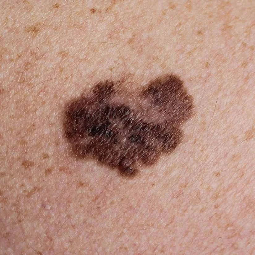
Melanoma The most dangerous form of skin cancer. Melanoma arises from the pigment-producing cells of the skin (melanocytes) and can spread to other organs if not caught early. It most commonly appears as a new or changing mole but can also arise in skin that looks normal. Early detection is critical — the five-year survival rate for melanoma caught at the earliest stage is over 98%, compared to significantly lower rates when detected late.
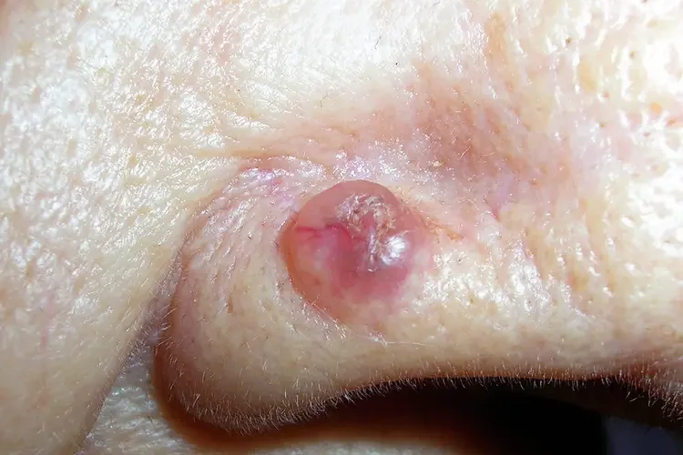
Basal Cell Carcinoma (BCC) The most common skin cancer in Australia. BCCs grow slowly and rarely spread to other parts of the body, but they can cause significant local damage if left untreated. They often appear as a pearly or pink nodule, a flat scar-like lesion, or a sore that doesn't heal. BCCs are almost always curable when treated early.
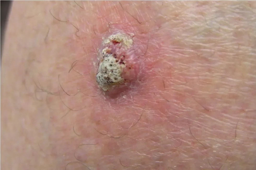
Squamous Cell Carcinoma (SCC) The second most common skin cancer. SCCs can grow more quickly than BCCs and carry a small but real risk of spreading if neglected. They typically appear as a thickened, scaly, or ulcerated lesion, often on sun-exposed areas. SCCs arising on the lip, ear, or in immunosuppressed patients tend to behave more aggressively and require prompt treatment.