Skin Biopsies
A skin biopsy is a minor medical procedure in which your doctor removes a small sample of skin tissue for laboratory examination under a microscope. It is one of the most reliable tools a doctor has for diagnosing skin conditions accurately.
Doctors perform skin biopsies for two main reasons:
To establish or confirm a diagnosis: particularly when the appearance of a skin lesion is uncertain, or when multiple diagnoses are possible.
To assess how well a treatment is working: by checking whether signs of disease are still present in the skin tissue.
Why has a biopsy been recommended for you?
Your doctor may have recommended a biopsy for one or more of the following reasons:
A spot on your skin looks unusual and a diagnosis cannot be made by visual examination alone
There are several possible diagnoses and the biopsy will help determine which is correct
A previously diagnosed skin condition needs to be monitored for activity or response to treatment
There is concern that a lesion could be cancerous or pre-cancerous
Types of biopsies:
There are several types of skin biopsy.
The two most common procedures performed in clinic are the shave biopsy and the punch biopsy. Your doctor will choose the most appropriate type based on the location, size, and nature of the skin concern.
Shave Biopsy
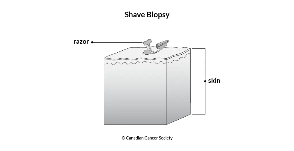
A shave biopsy involves using a thin, flexible blade to gently shave off the top layers of the skin (the epidermis and upper dermis). It is one of the most common biopsy techniques used in medicine.
-
Raised or superficial lesions, such as moles, warts, seborrheic keratoses, basal cell carcinomas, and other elevated skin growths
-
A small injection of local anaesthetic is given first to numb the area. You may feel a brief sting. Local anaesthetic takes away the feeling of pain but you can still feel pressure.
-
A sterile blade is used to slice across the skin surface to remove the lesion or a representative sample. No stitches are needed in most cases.
-
Typically 1–3 weeks. The area will form a scab and heal like a minor graze.
-
Usually minimal: a small, flat, pale mark. May be slightly lighter than surrounding skin.
Punch Biopsy
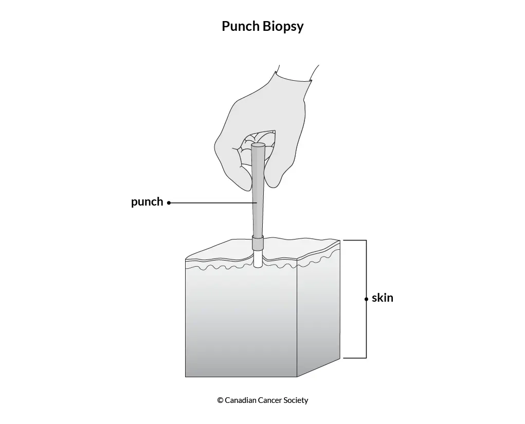
A punch biopsy uses a small circular blade (called a punch tool) to remove a cylindrical core of skin, going through the full thickness of the skin into the subcutaneous fat layer below. This provides a deeper tissue sample.
-
Flat or deep lesions: such as inflammatory conditions (eczema, psoriasis, lupus), rashes, pigmented lesions, suspected melanoma, or infections.
Suitable for any body area; particularly useful for deeper or flat lesions where the full skin thickness needs assessment
-
A small injection of local anaesthetic is given first. You will feel pressure but not pain during the procedure.
-
A circular punch (usually 2–6mm in diameter) is pressed into the skin and rotated to remove a small cylindrical plug. One or two stitches are usually placed to close the wound.
-
Stitches are removed after 5–14 days depending on the site. Full healing takes 2–4 weeks.
-
A small round or oval scar remains. Scarring is slightly more visible than a shave biopsy scar. Tattoos may be left distorted.